Fig. 2.
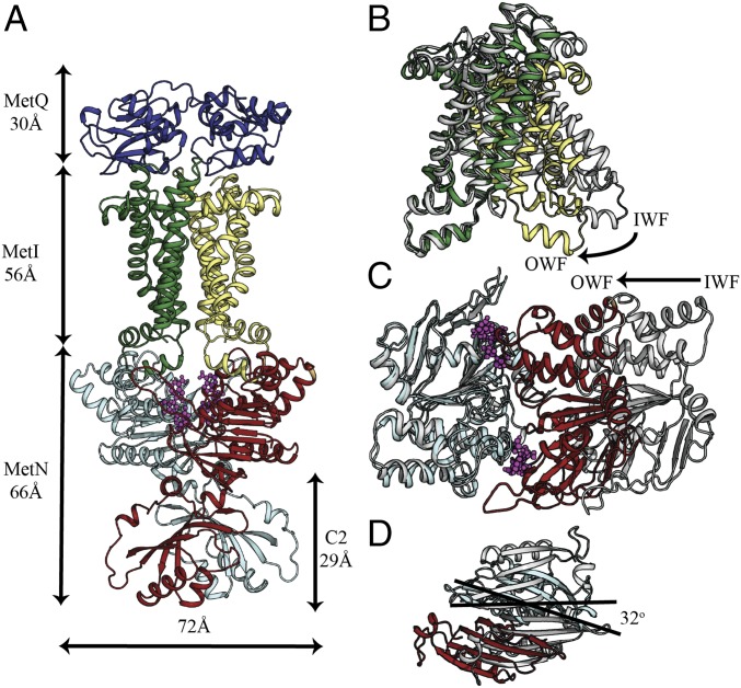
Crystal structure of the MetNIQ complex. (A) Side-view representation of the MetNIQ. MetQ subunit is colored slate, MetI subunits are forest and pale yellow, and MetN subunits are cyan and firebrick. C-regulatory (C2) domains are at the C termini of MetN subunits. Conformational changes of (B) the transmembrane MetI subunits, (C) the nucleotide-binding MetN subunits, and (D) the C2 domains between their OWF (colored as described in A) and IWF conformations (PDB ID code 3TUJ) (colored gray). One subunit of MetI (colored forest) or MetN (colored cyan) of the OWF conformation was overlaid to that of the IWF conformation to highlight the differences in the relative placement of the opposite subunit (B and C). Superposition of MetNIQ and MetNI reveals the rotation of the C2 domains around the molecular twofold axis between the two structures (D).

