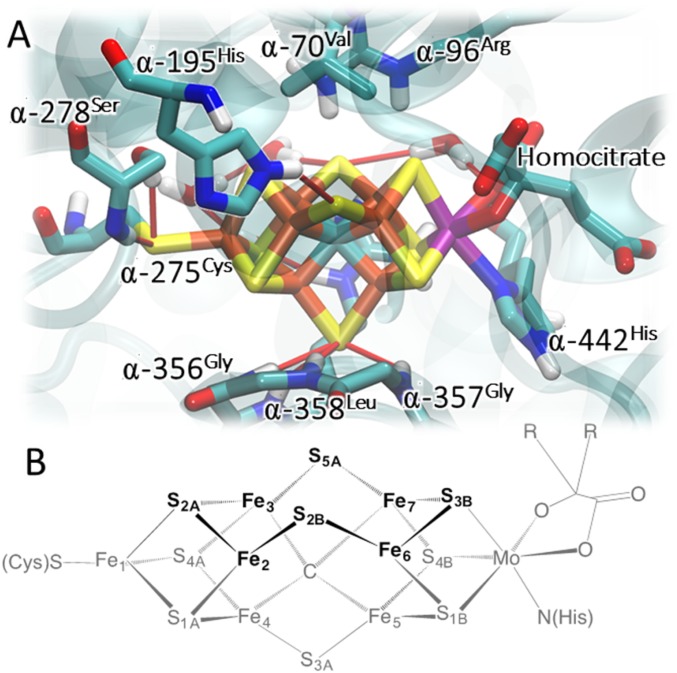
Fig. 1.
Structure of the FeMo-co binding pocket as obtained from MD simulations (22) (A) and schematic representation of FeMo-co (standard atom numbering) (B). For simplicity, only the polar H atoms are shown. The color coding of atoms is as follows: rust, Fe; yellow, S; cyan, C; purple, Mo; red, O; blue, N; light gray, H. Hydrogen bonds are shown as thin red sticks. The cartoon representation of the protein in cyan color is reported with the purpose to highlight the surrounding protein environment. The N2 access channel is indicated with the thick blue arrow.

