Fig. 2.
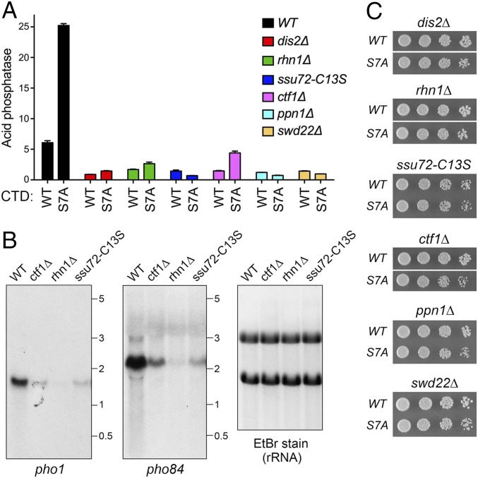
Role of termination factor Rhn1 and 3′-end–formation factors in pho1 expression in rpb1-CTD WT and CTD-S7A cells. (A) S. pombe cells bearing the indicated rpb1-CTD alleles were grown in liquid culture at 30 °C and assayed for acid phosphatase activity as described in Fig. 1C. Each datum in the bar graph is the average of assays using cells from at least three independent cultures ± SEM. (B) Northern blot analysis of total RNA from exponentially growing phosphate-replete WT, ctf1∆, rhn1∆, and ssu72-C13S cells. The RNA was resolved by agarose gel electrophoresis and stained with ethidium bromide to visualize 28S and 18S rRNA (Right) before transfer to membrane and hybridization with 32P-labeled probes specific for pho1 (Left) or pho84 (Middle). Annealed probes were visualized by autoradiography. The positions and sizes (in kilobases) of RNA size standards are indicated. The Northern blots shown are representative of three biological replicates of the experiment, entailing isolation and analysis of RNA from three separate cultures of each of the fission yeast strains specified. (C) Growth of dis2∆, rhn1∆, ssu72-C13S, ctf1∆, ppn1∆, and swd22∆ strains with rpb1-CTD-WT or rpb1-CTD-S7A alleles. Serial fivefold dilutions of exponentially growing cells were spotted on YES agar and then incubated at 30 °C. The pairs of WT and S7A strains were spotted on the same agar plate in every case. In the three instances shown in which the WT and S7A spottings are separated by a white space, other rows of cell spottings on the plate separating WT and S7A strains were cropped out of the image.

