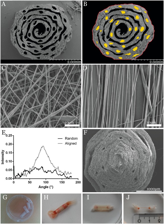
Figure 1.
PLC nerve guide characteristics. (A) SEM images of the PCL guide with more defined number of micro channels; (B) individual channel dimensions traced in ImageJ to calculate the porosity; (C) randomly oriented PLLA fibers; and (D) aligned PLLA fibers, indicating the porosity of the scaffold, and (E) the Fourier spectra of the random and aligned fibers. (F) SEM image of the PLLA nerve guide, where the pores appear to be less well defined than in the PCL guide; (G) the PCL nerve guide inserted in its silicone shell; (H) explanted nerve guide integrated in the regenerating sciatic nerve; (I) hollow tube bridged by a thin silicone matrix; (J) the regenerated matrix measured to the length of the sciatic nerve defect of 10 mm.

