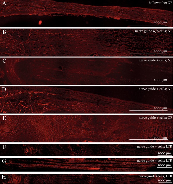
Figure 5.

Longitudinal sections of the PLLA nerve guide four weeks after sciatic injury and repair. Panel (A–E): Neurofilament (NF), ED1, and S-100 staining of (A) regeneration matrix in a hollow silicone tube; (B) nerve guide without cells; (C–E) and nerve guide with cells. Panel F–H: LipidTOX Red (LTR) staining of PLLA nerve guide + cells. Immunofluorescence microscopy rendered a faint background for both polymer types, which is due to light scattering in the fiber matrix and not due to autofluorescence.
