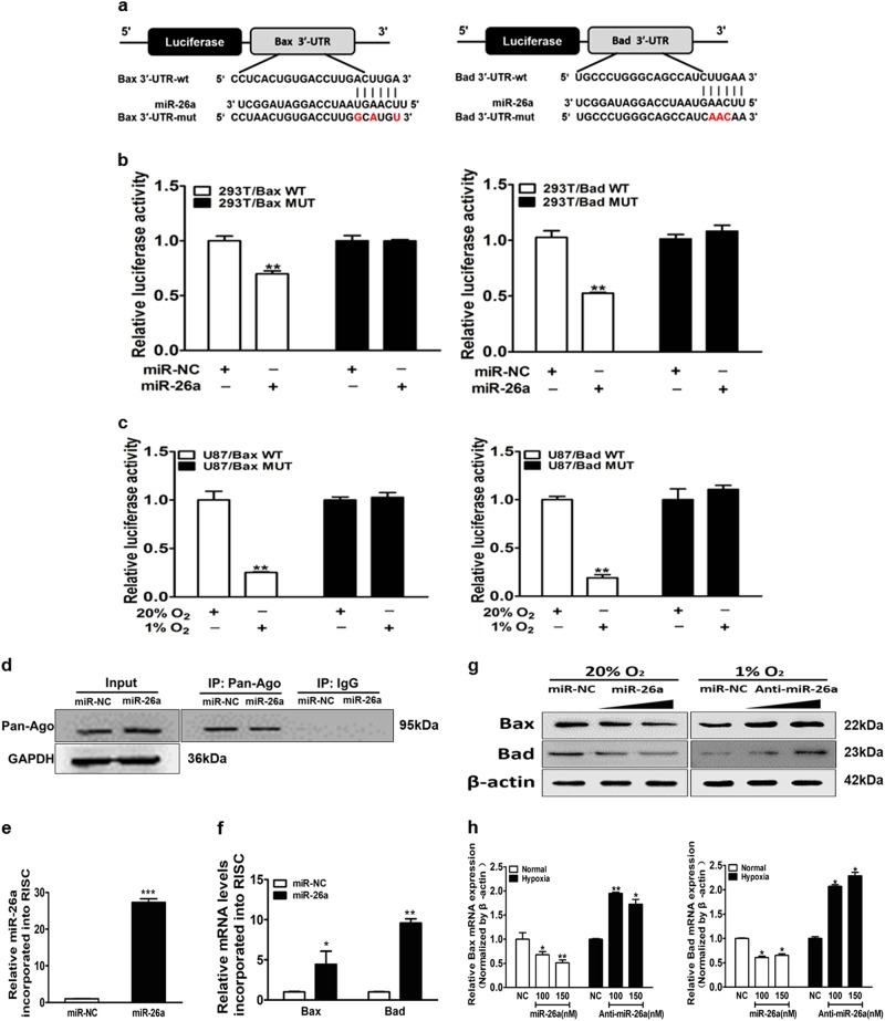
Fig. 4. Bax and Bad are direct targets of miR-26a.
a The complementary pairing of miR-26a with Bax and Bad wild-type (WT) and mutant (Mut) 3′-UTR reporter constructs were shown. b The reporter plasmids carrying the WT or Mut Bax and Bad 3′-UTR regions were co-transfected with miR-NC, miR-26a mimics and pRL-TK into 293T cells. After 24 h of the transfection, the relative luciferase/pRL-TK activities were analyzed. c WT or Mut 3′-UTR constructs of Bax and Bad were transfected into U87MG cells, then exposed to normoxia or hypoxia for 24 h. The relative luciferase/pRL-TK activities were determined. d Immunoblotting was used to detect the Ago2-RISC complex by the Ago2 antibody from U87MG cells with miR-NC or miR-26a mimics. Negative control was IgG. GAPDH was used as an internal control. e qRT-PCR was conducted to measure miR-26a levels incorporated into RISC in cells overexpressing miR-26a and miR-NC. U6 RNA levels were used as an internal control. f qRT-PCR analysis was performed to measure levels of indicated Bax and Bad mRNA levels incorporated into RISC derived from miR-26a or miR-NC cells. GAPDH levels were used as an internal control. g, h U87MG cells exposed to normoxia or hypoxia were transfected with miR-26a mimics (100 and 150 nM) or anti-miR-26a inhibitor (100 and 150 nM). Bax and Bad expression levels were determined by immunoblotting and qRT-PCR using β-actin as an internal control. Data are presented as mean ± SEM from three independent experiments. Asterisk indicates significant difference at p < 0.05 compared with control group and double asterisk indicates significant difference at p < 0.01 compared with control group

