
Figure 2. Current methods to visualize ceramide signaling.
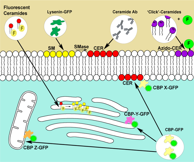
Fluorescent ceramides such as NBD-ceramides have been shown to accumulate in the Golgi apparatus and rapidly transform to NBD-sphingomyelin. Lysenin and other toxins have been shown to selectively bind sphingomyelin, recombinant expression fused to fluorescent proteins are used to visualize sphingomyelin (as ceramide precursor). Several ceramide antibodies have been shown to recognize ceramide. Generation and localization of ceramide in the plasma membrane have been demonstrated. Azido-ceramides and other functionalized ceramides can be combined with click chemistry to label ceramides with fluorophores in situ. Finally, several reported ceramide binding proteins have been shown to translocate to the plasma membrane to bind ceramide. This has been recognized by immunofluorescence or by fusion to the green fluorescent protein. Note that different CBP (X,Y,Z…) would recognize different pools of bioactive ceramide.