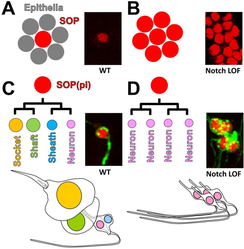
Figure 3: Phenotypic consequences of Notch signaling loss during mechanosensory organ development.
A) Notch signaling mediates the lateral inhibition to specify an SOP from a proneural cluster. Cells that receive high Notch signaling becomes epidermal cells. B) Upon loss of Notch signaling during lateral inhibition, all cells takes the SOP cell fate. Photographs show SOPs marked by Senseless expression (Red). C) Reiterative Notch signaling is required to specify the four cell fates of the mechanosensory organ. The cells that receives the highest amount of Notch signaling becomes the Socket cells while the cells that receive the least becomes the neuron. D) Upon loss of Notch signaling during lineage decisions all cells take on the neuronal fate. Photographs show neuronal nuclei and membrane, labeled by antibodies against Elav (Red) and Hrp (Green). Panels A and B were adapted and mofidied from [108].

