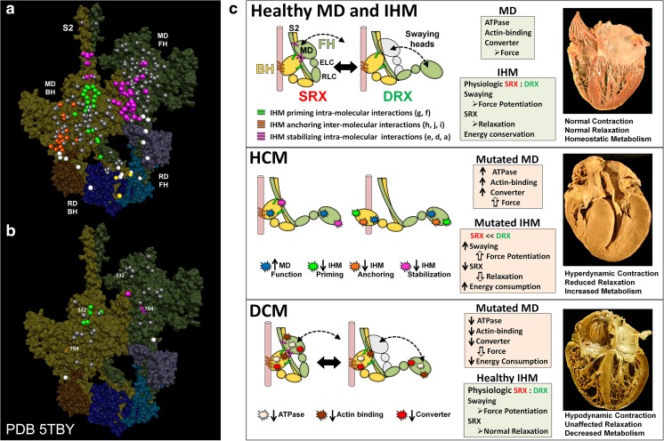
Fig. 4.
Assessment of the effects of hypertrophic (HCM) and dilated (DCM) variants according to their localization on the same positions on the blocked (BH) and free (FH) heads but exposed to different environments. Mapping of pathogenic and likely pathogenic HCM (a) and DCM (b) variants on the homologous human myosin β-cardiac IHM PDB 5TBY, based on the tarantula PDB 3JBH quasi-atomic model. Each variant appears as a pair, one located on or associated with the blocked head (olive) and one on the free head (green). Essential light chain ELC (BH, brown; FH, purple), regulatory light chain RLC (BH, dark blue; FH, light blue). a HCM variants that alter residues involved in IHM interactions (73/135 variants, 54%) are represented by colored balls: priming, green (“f” and “g”, see interaction colors code on c top panel); anchoring, orange (“i” and “j”); stabilizing, magenta (“a”, “d”, and “e”); scaffolding, white (ELC–MHC and RLC–MHC); RLC–RLC interface, yellow. Variants that do not alter residues involved in IHM interactions are shown in gray. b DCM variants (7/27; 26%) colored as described above. c The molecular pathogenesis of HCM and DCM assessed in the context of the IHM paradigm. Myosin interactions involved in IHM assembly and myosin motor domain (MD) functions that are altered by variants are depicted. Top panel: Relaxed healthy cardiac muscle contains myosin heads populations in the super-relaxed (SRX) state (left) with lowest ATP consumption and a disordered relaxed (DRX) state (right) with swaying free heads that generate force with higher ATP consumption (see Alamo et al. 2016). The population of cardiac myosins in SRX is more stable than in skeletal muscle (Hooijman et al. 2011), which supports physiologic contraction and relaxation, energy conservation, and normal cardiac morphology. Middle panel: HCM myosin variants (colored stars) both alter residues involved in MD functions (causing increased biophysical power, Tyska et al. 2000) and destabilize IHM interactions (particularly those with altered electrostatic charge). Reduced populations of myosins in the SRX state and increased populations of myosins in DRX as well as enhanced MD properties will result in increased contractility, decreased relaxation, and increased ATP consumption, the three major phenotypes observed in HCM hearts. Compensatory signals may promote ventricular hypertrophy. Bottom panel: MYH7 DCM variants (colored stars) have modest effects on IHM interactions but substantially reduce MD functions, particularly nucleotide binding, resulting in reduced ATP consumption and sarcomere power (Schmitt et al. 2006), with minimal impact on relaxation and overall diminished contractility. Compensatory signals result in ventricular dilatation to maintain circulatory demands in DCM hearts. Reproduced with permission from Alamo et al. (2017b) and eLife

