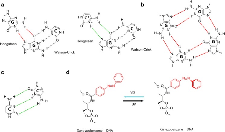
Fig. 3.
DNA structures that are sensitive to pH, ionic concentration and photoactivation. a Non-canonical Hoogsteen hydrogen-bonding patterns for G·G and G·C+ (red and green), shown beside the canonical Watson-Crick hydrogen bonding pattern of G·C (black). These form the basis of G-quadruplex and DNA triplex formation respectively. b The G-tetrad, formed by cyclic Hoogsteen-bonded (red) square planar alignments of four guanines that are stabilised by bound monovalent Na + and K+ cations (not shown). G-quadruplexes are formed by stacking of G-tetrads. c Semiprotonated C·C+ base pairs (green), which are intercalated to form the pH-sensitive i-motif. d trans-cis photoisomerization of azobenzene modification under UV and visible light (Vis) can be used to control DNA strand hybridization (trans) and dissociation (cis)

