
Fig. 1.
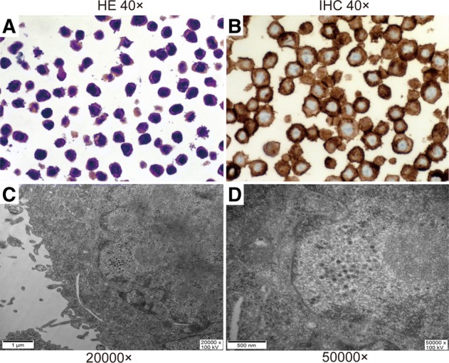
The identification of KGHV500 infected SW480 cells. (a) The HE staining of SW480 cells (40×). (b) The expression of CD46 on the surface of SW480 tumor cells is high and detected by immunohistochemistry staining. CD46’s and the main location is on the cell membrane (40×). (c and d) Many virus particles with a diameter of 70–90 nm in the cytoplasm and nucleus of SW480 cells (20000×) and (50000×) were detected by electron microscopy