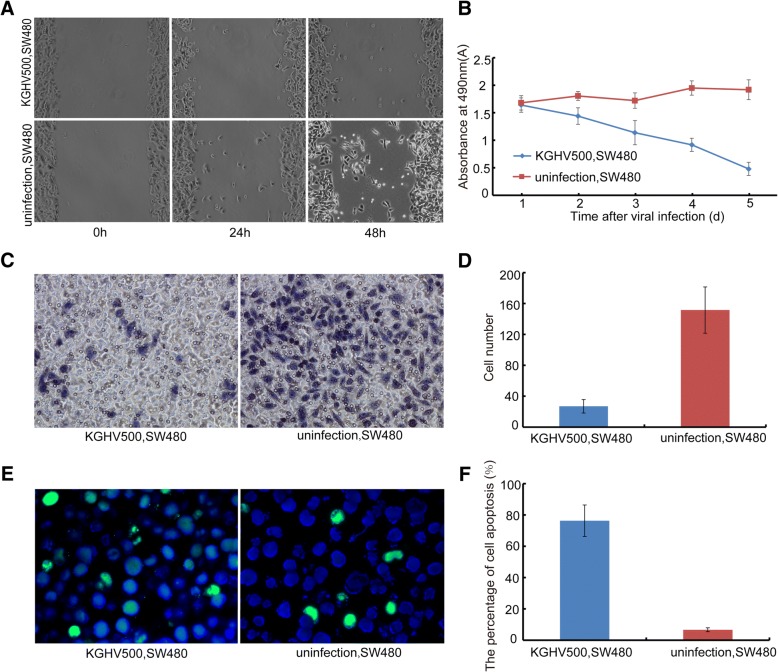
Fig. 2.
The anti-tumor efficacy of KGHV500 in vitro. (a) The result of the cell migration assay using SW480 cells infected by KGHV500 at an MOI of 100.0 for 0 h, 24 h, 48 h. The migration of SW480 cells was inhibited by KGHV500. (b) MTT assay of cell growth in SW480 cells treated with KGHV500 for 1d, 2d, 3d, 4d and 5d. The growth of SW480 cells was inhibited by KGHV500. (c and d) Transwell assay: The number of invading cells in the KGHV500-infected SW480 cells group was much lower than the number of invading SW480 cells in the control group (P < 0.01). (e) There were more apoptotic tumor cells found in the KGHV500 group by TUNEL staining (green: TUNEL, blue: DAPI). (f) The percentage of apoptotic cells was higher in the KGHV500 infected SW480 cells group than in the SW480 control group (P < 0.01). Data are presented as the mean ± s.d. *Significantly different from the control group (P < 0.05). **P < 0.01 vs controls

