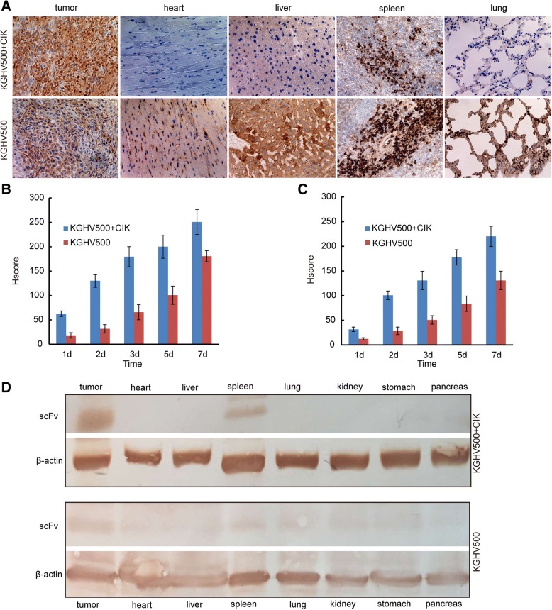
Fig. 5.
The expression of scFv and KGHV500 in xenograft tumors. (a) The expression of KGHV500 in xenograft tumor tissues (heart, liver, spleen and lung) was detected by immunohistochemistry after 7 days of treatment. There were more KGHV500 viruses in the KGHV500 combined with CIK cells treatment group. (b) The HSCORE of KGHV500, there were more positive cells in the KGHV500 combined with CIK cells group. (c) The HSCORE of scFv, there were more positive cells in the KGHV500 combined with CIK cells group. (d) The expression of scFv in organs (heart, liver, spleen, lung, kidney, stomach and pancreas) of nude mice treated with KGHV500 combined with CIK cells and KGHV500 was detected by Western blot. scFv was expressed only in the tumor and spleen in the KGHV500 combined with CIK cell treatment group, but expression of scFv was found in all tissues in the KGHV500 treatment group

