
Fig. 2.
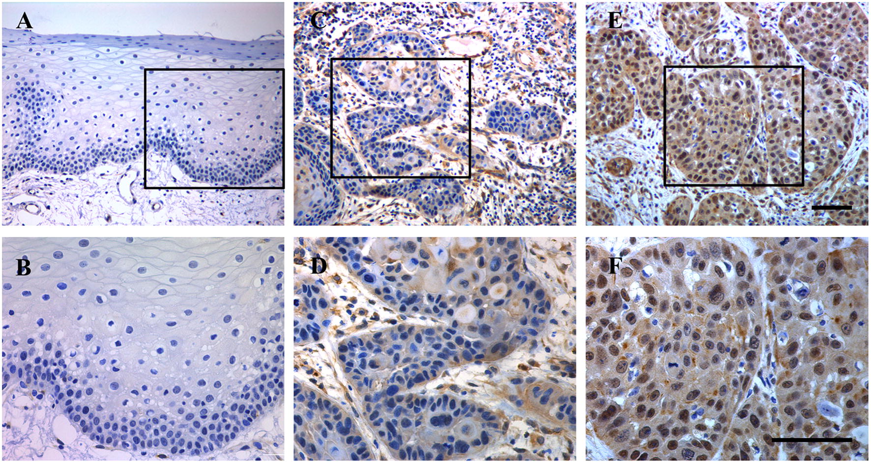
Immunohistochemical staining of TEAD4 in human HNSCC samples. A, B Representative negative staining of TEAD4 in normal oral epithelial; C, D representative low expression of TEAD4 in primary human HNSCC sample; E, F representative high expression of TEAD4 in primary human HNSCC sample. Nuclei are counterstained with hematoxylin. The areas marked by black box in the A, C, E images (upper panel) were shown in larger magnification as B, D, F images (lower panel), respectively. Scale bar: 100 μm