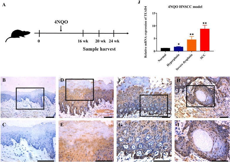
Fig. 4.
TEAD4 expression pattern during HNSCC tumorigenesis in 4NQO-induced animal model. A Experimental scheme of 4NQO-induced HNSCC animal model. B–I Immunohistochemical staining of TEAD4 in samples from diverse stages in 4NQO-induced animal model. Images in the upper panel (B, D, F, H) were representative staining of TEAD4 in normal, epithelial with hyperplasia, epithelial with severe dysplasia/carcinoma in situ and squamous cell carcinoma, respectively. Images in the lower panel (C, E, G, I) were magnified from the black box area in the B, D, F, H images in the upper panel, respectively. Scale bar: 100 μm. J The mRNA levels of TEAD4 during the 4NQO-induced HNSCC were measured by qRT-PCR in pre-stored samples (n = 5, 6 samples per group). *P < 0.05, **P < 0.01, ANOVA analyses

