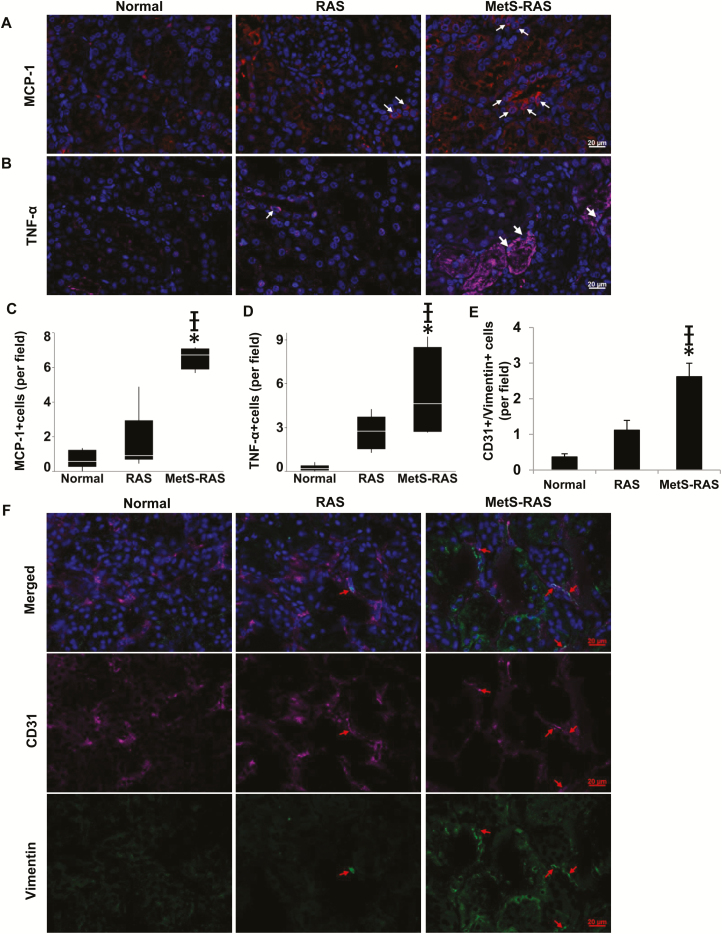
Figure 4.
In immunofluorescence, MCP-1-expressing cells (red) were more abundant in MetS-RAS compared with normal and RAS (a, c), as was TNF-α expression (pink) (b, d). CD31 (pink) and Vimentin (green) colocalization was higher in MetS-RAS than in RAS and normal, suggesting increased endothelial to mesenchymal transition (e, f). (Please refer to color online only for indicated colors). *P < 0.05 vs. normal, †P < 0.05 vs. RAS. Abbreviations: MCP-1, monocyte chemoattractant protein-1; MetS-RAS, metabolic syndrome and renal artery stenosis; RAS, renal artery stenosis; TNF-α, tumor necrosis factor-alpha.

