Figure 1.
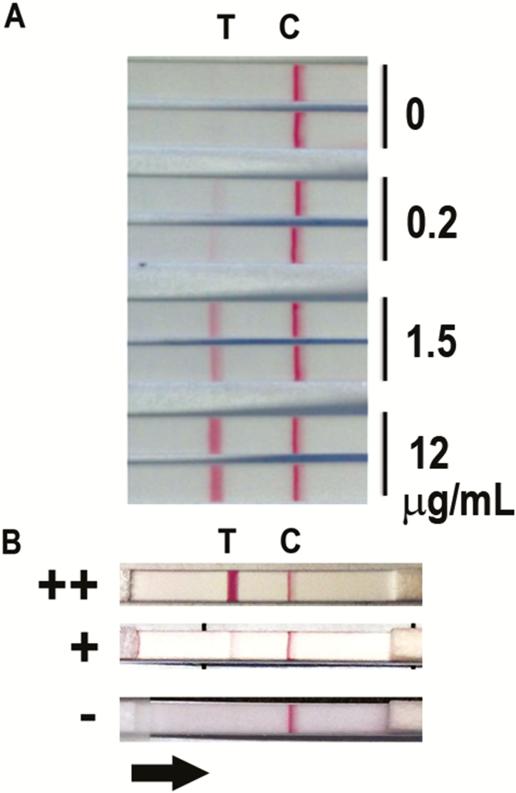
A, Representative prototype dipstick positivity at variable concentrations (0, 0.2, 1.5, and 12 µg/mL) of ethanol precipitate antigen spiked into healthy human urine. Two replicates are shown for each antigen condition. Line “C” represents the position of the control line containing control antibody and line “T” represents the position of β-galactofuranose-recognizing mAb476 on the membrane. Binding of Aspergillus antigen within ethanol precipitate or sample marks the T-line. B, Representative test results of clinical samples showing semiquantitative visual appearance of negative (–), low-positive (+), and high-positive (++) interpretations. The black arrow represents the direction of flow of the sample in the assay.

