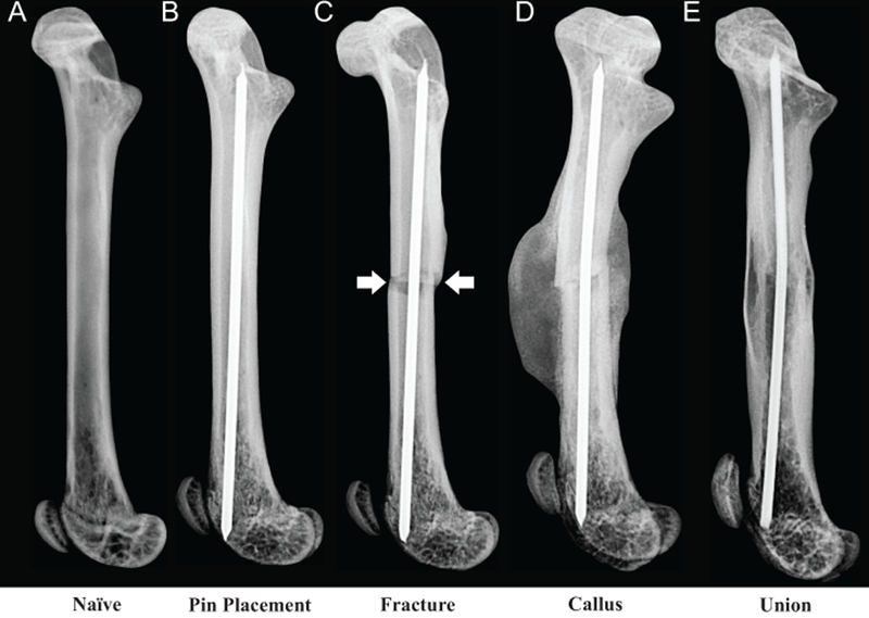
Figure 1. Radiographic images of the mouse model of bone fracture pain.

A 0.5-mm hole was drilled in the center of the trochlear groove of naïve C57Bl/6J male mice (A), 8 to 9 weeks old. A pin was then placed (B) into the intramedullary canal, the drill site was sealed with a dental amalgam plug and the mice were allowed to recover for 3 weeks before pre-fracture activity was measured. A closed mid-diaphyseal fracture (arrows) of the femur (C) was performed using a 3-point impact device. Formation of a large mineralized callus (D) occurs at 14-days post-fracture and union of the cortical bone (E) is apparent at day 63 post-fracture.
