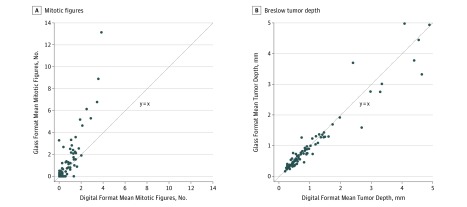
Figure 3. Case-Level Comparisons of Interpretive Features Using Traditional vs Digital Format.
These figures show that there is a higher number of mitotic figures seen using traditional microscopy but no difference in Breslow tumor depth. A, The 63 points represent the 87 cases included; 21 cases had 0 mitotic rate for all traditional and digital interpretations, and 4 other points represent 2 cases each. B, The 87 points represent the 87 cases included. All traditional-digital combinations of mean case-level Breslow depth are distinct.

