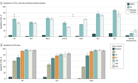
Figure 1. Gentian Violet (GV)–Induced Apoptosis in Cutaneous T-Cell Lymphoma Cells.
Cells were treated with GV, nitrogen mustard (mechlorethamine), or dimethyl sulfoxide (DMSO) at 1 μmol/L for 24 hours; apoptosis was detected by flow cytometry. The Jurkat T-cell acute lymphoblastic luekemia cell line line was included for comparison. A, P < .01 for GV compared with DMSO for all. B, P < .01 between each GV-treated column with the DMSO-treated column. SS1 indicates Sézary syndrome 1; SS2, Sézary syndrome 2.
aP < .01 compared with nitrogen mustard.

