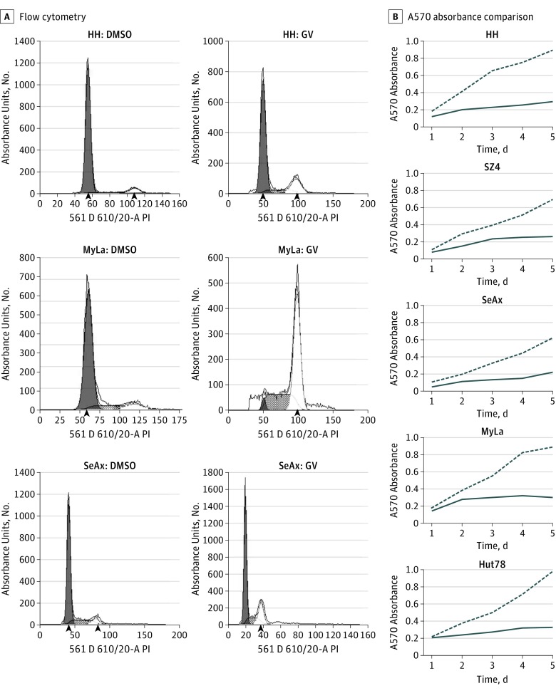
Figure 4. Gentian Violet (CV) Inhibition of Cell Proliferation and Induction of Cell Cycle Arrest at G2/S Phases in Cutaneous T-Cell Lymphoma Lines.
Cells were treated with GV (1 μmol/L) or dimethyl sulfoxide (DMSO) (1 μmol/L). Cell cycle and proliferation were measured by propidium iodide staining and flow cytometry and MTT assay, respectively. A, Flow cytometry plots show cell cycle arrest after GV treatment (48 hours). Arrowheads indicate the the median fluorescence intensity of the G0/G1 and G2 populations. B, A570 absorbance comparison of GV- and DMSO-treated cells at days 1 to 5 shows decreased cell proliferation.

