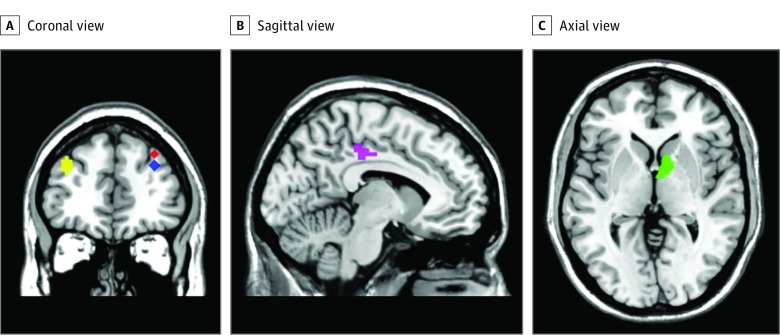
Figure 1. Regions of Interest Showing Differences in Brain Activation Between Low (L) and High (H) Psychoticlike Experiences Groups.
Coronal (A), sagittal (B), and axial (C) views. At age 14 years: right middle frontal gyrus (33, 41, 40 [red]) (L>H [low group has shown greater brain activation than high group]); right middle frontal gyrus (33, 44, 31 [blue]) (L>H); left cingulate gyrus (−12, −28, 40 [purple]) (L>H); and left middle frontal gyrus (L>H) (−36, 47, 31 [yellow]). At age 19 years: (9, 8, 1 [green]) right caudate head (L>H).

