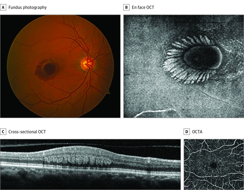
Figure 1. Case 1.
A, Fundus photography of the right eye at presentation displayed central macular hemorrhage in a characteristic radial pattern. B, En face optical coherence tomography (OCT) of the right eye at presentation illustrated radial hyperreflectivity at the level of the outer plexiform layer of Henle. C, Cross-sectional OCT of the right eye displayed intraretinal hemorrhage radiating in the outer plexiform layer of Henle. D, At the 6-week follow-up visit, there was remarkable improvement of the radial hemorrhage, and OCT angiography (OCTA) of the deep retinal capillary plexus illustrated microvascular abnormalities consistent with macular telangiectasia type 2.

