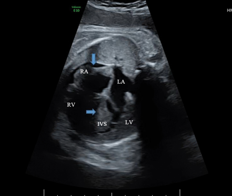
Fig. 1.

Fetal echocardiogram demonstrating severe biventricular hypertrophy and asymmetric septal hypertrophy demonstrated by
 . The right atrial wall is also noted to be hypertrophied indicated by
 . A small pericardial effusion is noted along the right ventricular side. IVS, interventricular septum; LA, left atrium; LV, left ventricle; RA, right ventricle; RV, right ventricle.
