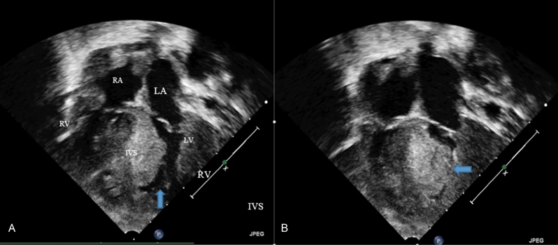
Fig. 3.

A postnatal echocardiogram showing severe HCM. (
A
) An end-diastolic still frame showing severe hypertrophy and a small end-diastolic ventricular cavity as marked by
 (
B
) An end-systolic still frame showing complete obliteration of the ventricular cavity in systole as shown by
 . IVS, interventricular septum; LA, left atrium; LV, left ventricle; LV, left ventricle; RA, right atrium.
