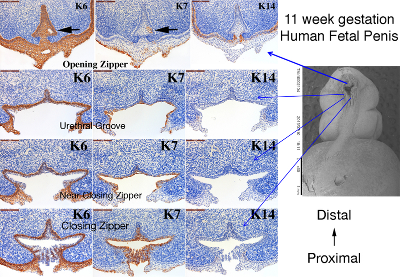
Figure 10.
Cytokeratin (K) 6, 7 and 14 expression in an 11-week human fetal penis. Representative sections from the opening zipper, urethral groove, near the closing zipper and the closing zipper. Approximate section location is depicted by the blue arrows in the scanning electron microscopic image. Note the canalization (black arrows) in the opening zipper. Note the K6 localization to basal epithelial cells and K7 apical epithelial cells, and the complex arrangement of epithelial cells fusing in the closing zipper.

