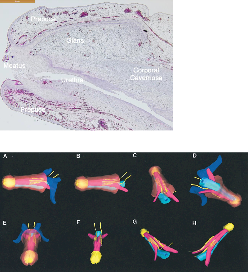
Figure 4:
Optical projection tomography of male urethral development from 6.5 to 10.5 weeks fetal age. Note the urethral plate (blue arrow) that ends within the glans. The wide-open urethral groove (red arrows) is best seen from 9.5 to 10.5 weeks with clear progression of proximal to distal fusion of the edges of the urethral groove to form the tubular urethra (yellow arrow). The epithelial tag is marked by the light blue arrow. The proliferation marker Ki67 are labeled with arrows in the OPT specimens A-G, with the exception of C which illustrates no staining for the apoptotic marker caspase 3. Canalization of the urethral plate is visible in histologic section A-D.
Reproduced from Li, Y., A. Sinclair, M. Cao, et al., Canalization of the urethral plate precedes fusion of the urethral folds during male penile urethral development: the double zipper hypothesis. J Urol, 2015. 193(4): p. 1353–59, with permission.

