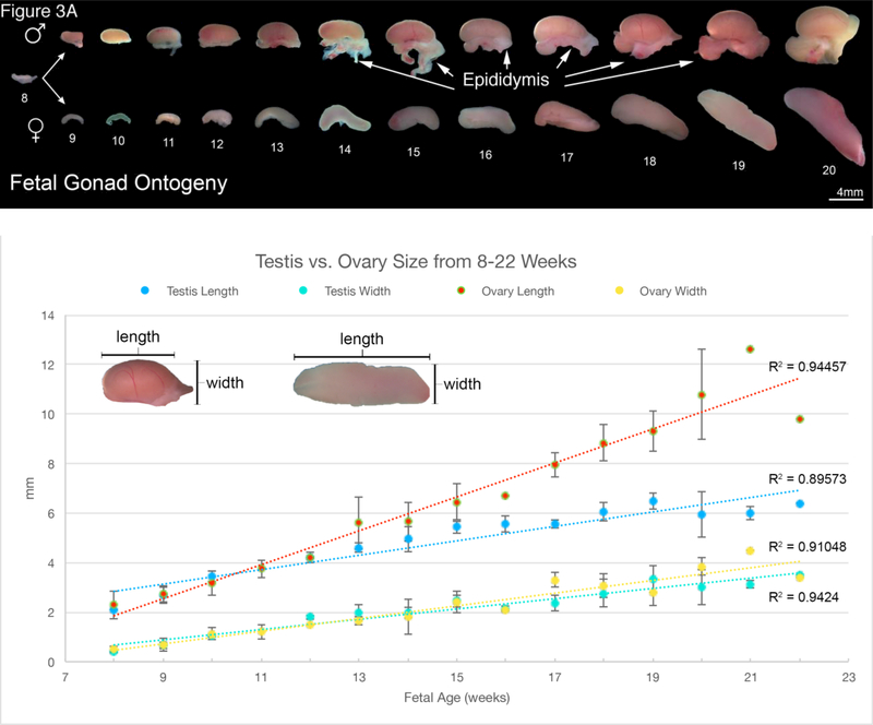
Figure 3.
A. Wholemount photos of human fetal gonadal ontogeny. Testis ontogeny is on the top row and ovarian ontogeny on the bottom row. The testis has a more spheroid shape in contrast to the oblong shape of the ovary. Note the epididymis in many of the testicular specimens. B. Morphometrics (in millimeters) of the human testis and ovary during fetal development (weeks).

