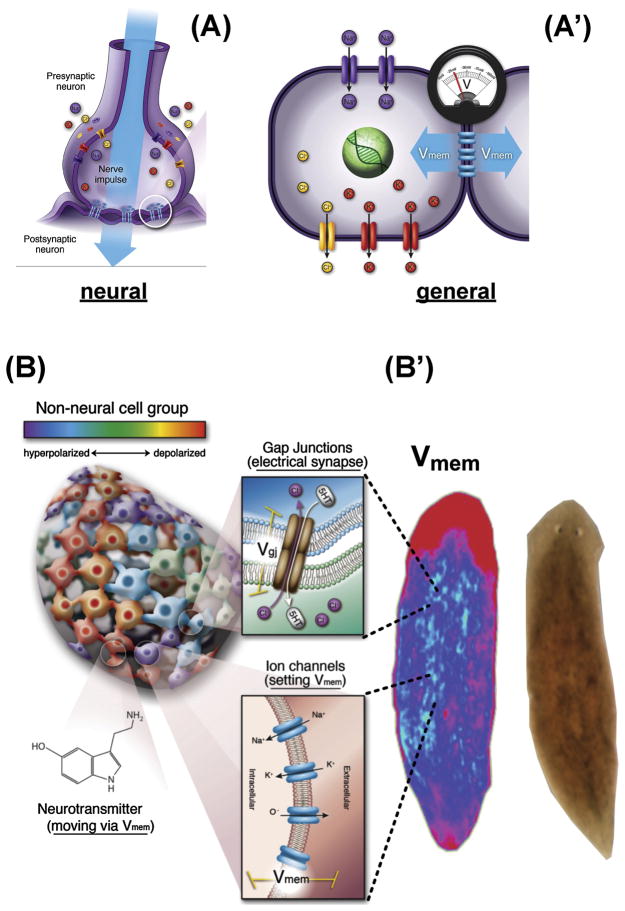
Figure 2. Bioelectric signaling among somatic tissues.
(A) Neurons implement memory and distributed decision-making by virtue of electrical potentials (Vmem) set by ion channels, which are propagated to neighboring cells via electrochemical synapses known as gap junctions.
(A′) The same machinery is present in most cells, where ion channels and pumps set Vmem, and gap junctions allow its propagation to some neighboring cells.
(B) Tissues sustain physiological compartments, whose borders and patterns of small molecule connectivity that are driven by the complex gating of ion channels and gap junctions. As in the central nervous system, neurotransmitters are among the key small molecule morphogens moved across tissues by bioelectric properties.
(B′) These dynamics result in spatio-temporal distributions of resting potential across anatomical distances (shown here in a planarian) – bioelectrical prepatterns that underlie subsequent gene expression and other cell behaviors during regeneration and development.
Panels A, A′ were created by Jeremy Guay of Peregrine Creative.

