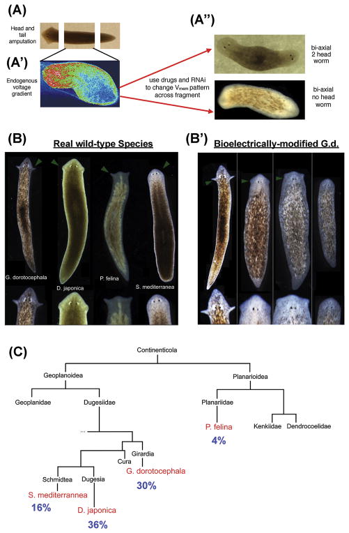
Figure 3. Bioelectrically-mediated changes of patterning in planaria.
(A) D. japonica mid-fragments exhibit bioelectric gradients, with anterior ends’ cellular Vmem depolarized compared to those of posterior cells. This pattern can be detected [85] via voltage-sensitive fluorescent dyes (A′), and modified with ion channel drugs, which alter the endogenous bioelectrical gradient toward bi- or no-head heteromorphoses respectively (A″), demonstrating that the bioelectric pattern is instructive for large-scale anatomical polarity along the AP axis [100, 132].
(B) G. dorotocephala planaria exhibit a characteristically distinct head shape, compared to other species. (B′) When fragments of G. dorotocephala were briefly treated with a gap junction blocker [124], they regenerated heads whose shapes matched those of other extant species of planaria.
(C) The appearance of these head shapes in a single cohort of worms treated together was stochastic, appearing at frequencies proportional to the evolutionary distance between the species.
Panel A′ was courtesy of Taisaku Nogi. Panels B–C used with permission from [124].

