. 2018 Sep 14;48(6):377–383. doi: 10.4041/kjod.2018.48.6.377

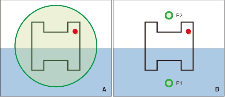
Figure 1. Bovine enamel blocks with bonded orthodontic brackets (red dot indicates the distogingival aspect) prepared for colorimetric evaluation of white spot lesions following external bleaching with or without fluoridation. The blue area describes the area covered with nail varnish. The brackets and nail varnish were absent at the time of spectroradiometer analysis and digital image acquisition. A, The green circle represents the area of measurement by the spectroradiometer. B, The green circles (P1, sound enamel area; P2, white spot lesion area) describe the areas of measurements by Photoshop after the acquisition of digital images.

Figure 1