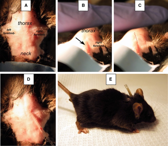
Figure 2.

Landmarks used to predict the incision point on the skin above the right external jugular vein of a C57Bl/6 mouse. Panel A shows the surgical area prior to catheter placement with anatomical landmarks indicated. Panel B shows the skin infolding (arrow) used to identify the location of the jugular vein. Panel C shows the approximate path of the jugular vein, indicated by a dotted line. However, the neck shape and the appearance of skin infoldings vary with mouse background strain. Panel D shows how the optimal incision point is located by the indelible mark in the catheter that is placed above the infolding. Panel E shows a catheterized mouse with the capped catheter protruding from its back.
