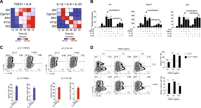
Fig. 1. AHR is activated during in vitro pathogenic TH17 cell differentiation, and it regulates IL-17A production in a TGFβ1-dependent manner.
a Heatmap of Ahr, Cyp1a1, Ahrr, Il22, and Il17a mRNA expression in CD4+CD44loCD62Lhi naive T cells differentiated for 12, 24, 36, 48, 60, and 72 h under nonpathogenic (TGFβ1 plus IL-6) and pathogenic (IL-1β, IL-6, and IL-23) TH17 conditions. b qPCR analysis of Ahr, Cyp1a1, and Ahrr mRNA expression of naive CD4+ T cells (white bars) activated 24 h under different combinations of IL-6, TGFβ1, IL-1β, and IL-23 (as indicated). c Frequency of IL-17A+ and IL-22+ cells from pathogenic TH17 cells differentiated with TGFβ1, IL-6 and IL-23 (pTH17 (TGFβ1)) or IL-1β, IL-6 plus IL-23 (pTH17 (IL-1β)) in the presence of FICZ. d Frequency of IL-17A+ and IL-22+ cells from pTH17 cells (TGFβ1, IL-6, and IL-23) differentiated under different concentrations of TGFβ1. b P < 0.05 when compared to amedium; bIL-6; cTGFβ1, and dTGFβ1+IL-6 groups (two-way ANOVA). NS not significant; *P < 0.05, *P < 0.01, and ***P < 0.001 (c, unpaired, two-tailed Student’s t test and d two-way ANOVA). Data are representative of more than three independent experiments with similar results

