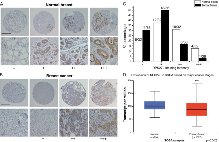
Fig. 6. RPS27L levels were reduced in human breast cancer tissues.
a, b RPS27L staining in normal breast tissues (a) and breast cancer tissues (b). Breast tissue microarrays containing normal breast and tumor tissues were stained for RPS27L expression. Stained normal and tumor tissues were classified into four groups (− to +++) according to the staining intensity. Scale bar in top panels = 500 μm, scale bar in bottom panels = 100 μm. c The percentage of normal or tumor tissues in each staining group. Tissue samples with different staining intensity were grouped and indicated. d RPS27L transcript was significantly reduced in primary breast tumors from TCGA database. p = 0.002

