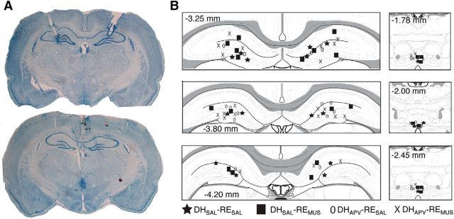
Figure 5.
Cannula placements in the RE and DH. A, Representative thionin-stained coronal sections showing a midline cannula placement in the RE (left) and DH (right). B, Cannula placements for all subjects that were included in the analysis across three different levels along the anterior–posterior axis in both DH (top) and RE (bottom). The distribution of cannula placements within the RE and DH was similar across all of the groups.

