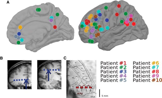
Figure 1.
Bipolar SEEG recordings obtained from epileptic patients. A, Locations of 60 cortical bipolar recordings from 10 patients with intracranial electrodes. B, Illustration of bipolar transcortical derivation used throughout the study, with arrows indicating two cortical bipolar channels from Patient 3. C, Illustration of bipolar recordings from Patient 1 from the pulvinar of the thalamus.

