Abstract
The bench press (BP) is a complex upper body exercise in which substantial external loads can be used, demanding high neuromuscular activity. The aim of this study was to compare electromyographic (EMG) activity between female and male athletes during the flat bench press. Five male and five female athletes participated in this study. The main session included four sets of one repetition of the flat bench press with the load of 55, 70, 85 and 100% of the one-repetition maximum (1RM). The activity of four muscles was analysed: the pectoralis major (PM), the anterior deltoid (AD), the lateral head of the triceps brachii (TBlat) and the long head of the triceps brachii (TBlong). The main finding of the study was that the muscle activity pattern differed between women and men during the bench press depending on the external load. The non-parametric Kruskal-Wallis ANOVA for males showed differences between the TLpeak values recorded for different loads (55%-100% 1RM) during the bench press (chi-square = 15.3, p = 0.009) and ADpeak (chi-square = 19.5, p = 0.001). The non-parametric Kruskal-Wallis ANOVA for females showed differences between the ADpeak values recorded for different loads (55%-100% 1RM) during the bench press (chi-square = 12.1, p = 0.018).
Keywords: Bench press, EMG, Resistance exercise, Testing
INTRODUCTION
Training to increase neuromuscular fitness has been shown to be effective in improving athletic performance [1]. Neuromuscular training consists of overcoming external resistance using internal strength, with multiple sources of resistance available, such as dumbbells, barbells, as well as elastic, pneumatic and hydraulic resistance [2]. The bench press (BP) is a complex upper body exercise in which substantial external loads can be used, demanding high neuromuscular activity. The potential of the BP for strength development and popularity of BP competitions have made it a unique phenomenon as a popular exercise for training, testing or research purposes. Athletes are often involved in specific training programmes that can change the proportions of strength in different muscle groups. Interesting insight into these problems can be derived from the topography of muscle strength that describes how particular groups of muscles contribute to total strength. Scientists and coaches are interested in how maximum strength, explosive strength or power output can be improved using the BP exercise and how muscle activity changes for BP variations [3].
The neuromuscular recruitment has been assessed only by the EMG amplitude to observe the influence of exercise load as an external stimulus to provide a greater training stimulus to muscles. That is, only the tonic aspect of neurophysiologic behaviour of motor units during muscular contraction related to intensity of muscular activation has been considered. Two previous reviews have been written to answer these questions. The first review evaluated the criteria for BP efficiency and safety that can be recommended for strength conditioning programmes [4, 5], while the second one provided a meta-analysis of the studies focused on optimal load for power training [6, 7].
Depending on the method used to perform the bench press exercise, the following three muscle groups are primarily involved: the pectoralis major (PM), the anterior deltoid (D) and the triceps brachii (TB) [8, 9]. The change in the load affects the pattern of muscle activity during this exercise [10]. The root mean square (RMS) value provides information about the geometric (amplitude) and temporal characteristics of motor unit behaviour during muscular contraction. It is important to understand the tonic and phasic characteristics of neuromuscular stimuli to control the performance under different external loads. Although internal movement structure has been described extensively in the literature concerning the bench press [11,12,13], no studies have compared muscle activity patterns between female and male athletes. The main aim of this study was to compare the muscular activity patterns between male and female athletes during the flat bench press. The specific aims were to evaluate differences in muscular activity patterns between female and male athletes at various external loads during the bench press and to determine the muscle groups which show the most significant differences between female and male athletes.
MATERIALS AND METHODS
Participants
Five male (age: 21 years; body height: 177 ± 8 cm, body mass: 85 ± 11 kg, 1RM bench press: 105 15 kg) and five female (age: 21 years; body height: 165 ± 8 cm; body mass: 69.7 ± 5.9 kg, 1RM bench press: 55 ± 10 kg) athletes with at least one year of bench press training experience participated in the study. The participants did not perform any resistance training 72 hours prior to testing to avoid fatigue. All the subjects were informed verbally and in writing about the procedures, as well as the possible benefits and risks of the tests and provided written consent before they were included in the study. The study received the approval of the Bioethics Committee at the Academy of Physical Education in Katowice, Poland.
Procedures
The measurements were performed in the Strength and Power Laboratory at the Academy of Physical Education in Katowice. There were two sessions of the experiment. A standardized warm-up protocol was used for each session, including a general warm-up (5 min) performed on a hand cycle ergometer (heart rate 130-140 bpm). The specific part of the warm-up consisted of three bench press sets with the load adjusted to perform 15, 10 and 5 repetitions. The first session was aimed at determination of the one-repetition maximum in the flat bench press (1RM). The second session included four sets of one repetition of the flat bench press with the load of 55, 70, 85 and 100% of 1RM. The activity of four muscles was analysed: the pectoralis major (PM), the anterior deltoid (AD), the lateral head of the triceps brachii (TBlat) and the long head of the triceps brachii (TBlong).
Measurements
An eight-channel Noraxon TeleMyo 2400 system (Noraxon USA Inc., Scottsdale, AZ; 1500 Hz) was used for recording and analysis of electric potentials from the muscles. The activity was recorded for four muscles: the pectoralis major (sternocostal fibres), the anterior deltoid, the triceps brachii (lateral head) and the triceps brachii (long head). Before placing the gel-coated self-adhesive electrodes (Dri-Stick Silver circular sEMG Electrodes AE-131, NeuroDyne Medical, USA), the skin was shaved, abraded and washed with alcohol. The electrodes (11 mm contact diameter and a 2 cm centre-to-centre distance) were placed along the presumed direction of the underlying muscle fibres according to the recommendations of Seniam [14]. The grounding electrode was placed on the connection with the triceps brachii muscle. Video recording was used for identification of the beginning and completion of the movement. The rate of all exercises was controlled by an electronic metronome (Korg MA-30, Korg, Melville, New York, USA). Analysis was based on peak muscular activity during the BP (both from the eccentric and concentric phases).
Statistical Analysis
Data normality was tested with the Shapiro-Wilk test. Changes in the activity of the muscles during the flat bench press for different loads were analysed using the Kruskal-Wallis non-parametric ANOVA [15]. Post-hoc tests were used to analyse statistically significant changes. Fixed-base indices were employed to evaluate changes in muscle activity. All statistical analyses were conducted by means of the STATISTICA 9.1 package and MS Excel 2010.
RESULTS
The non-parametric Kruskal-Wallis ANOVA for females showed differences between the ADpeak values recorded for different loads during the bench press (chi-square = 12.1, p = 0.018). As shown by the results of the post-hoc tests, the greatest differences occurred for the load of 55% of 1RM and 100% of 1RM (p = 0.044).
The non-parametric Kruskal-Wallis ANOVA for male athletes showed differences between the TLpeak values recorded for different loads during the bench press (chi-square = 15.3, p = 0.009). As shown by the results of the post-hoc tests, the greatest differences occurred for the load of 55% of 1RM and 100% of 1RM (p = 0.033). Variations were also found in the ADpeak values recorded for different loads during the bench press (chi-square = 19.5, p = 0.001). As shown by the results of the post-hoc tests, the greatest differences occurred for the load of 55% of 1RM and 100% of 1RM (p = 0.021) (Table 1, Figure 1, Figure 2).
TABLE 1.
Peak muscle activity for the PM, AD, TBlatpeak and TBlongpeak muscles during the bench press exercise performed by female and male athletes with the load of 55%, 70%, 85% and 100% 1RM.
| 55% 1RM | 70% 1RM | 85% 1RM | 100% 1RM | |
|---|---|---|---|---|
| FEMALES | ||||
| ADpeak | 90 | 119 | 125 | 151 |
| PMpeak | 65 | 67 | 85 | 95 |
| TBlatpeak | 51 | 55 | 67 | 68 |
| TBlongpeak | 55 | 59 | 62 | 75 |
| MALES | ||||
| ADpeak | 67 | 79 | 90 | 117 |
| PMpeak | 70 | 72 | 90 | 89 |
| TBlatpeak | 65 | 71 | 85 | 89 |
| TBlongpeak | 95 | 124 | 141 | 165 |
Note: the one-repetition maximum - 1RM; the pectoralis major - (PM); the anterior deltoid - (AD); the lateral head of the triceps brachii - (TBlat); the long head of the triceps brachii - (TBlong).
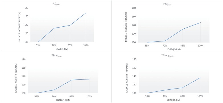
FIG. 1.
Muscle activity index (%) for ADpeak, PMpeak, TBlatpeak, TBlongpeak by female athletes during the flat bench press with a load of 55%, 70%, 85% and 100% 1RM.
Note: the pectoralis major - (PM); the anterior deltoid - (AD); the lateral head of the triceps brachii - (TBlat); the long head of the triceps brachii - (TBlong).
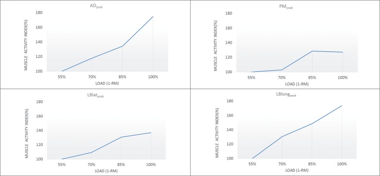
FIG. 2.
Muscle activity index (%) for ADpeak, PMpeak, TBlatpeak, TBlongpeak by male athletes during the bench press with a load of 55%, 70%, 85% and 100% 1RM.
Note: the pectoralis major - (PM); the anterior deltoid - (AD); the lateral head of the triceps brachii - (TBlat); the long head of the triceps brachii - (TBlong).
DISCUSSION
The aim of this study was to compare electromyographic activity between female and male athletes during the flat bench press. The main finding of the study is that the muscle activity pattern differs between women and men during the bench press depending on the external load. The electromyographic data determine whether the muscle is active, the level of muscle activity, how the muscles work together and whether muscular fatigue occurs [16]. The main aim of the study was to identify the pattern, i.e. to determine the muscle activity expressed by the percentage contribution of the activity to the specific exercise.
According to Sakamoto and Sinclair [11], once a strategy set by the central nervous system to perform a motor task is chosen, it is implemented by activation of a group of muscles in the appropriate sequence. The selection of the correct muscles to be activated is simplified by certain principles [17, 18]. One of these principles is directed at optimizing muscle coordination in order to minimize energy expenditure. Another principle is related to the prediction of forces such as gravity or inertial interactions among body segments [19]. The motor unit recruitment and firing rate to execute intended movements are regulated by the descending command from the central nervous system and can be modulated by afferent feedback during muscular weakness or fatigue [20]. The activity was recorded for the most important muscles of the shoulder girdle [21] involved in the bench press. These include the pectoralis major (the sternal head), deltoid (anterior head) and triceps brachii (the lateral and long head). Numerous authors [22, 23, 24, 25] have confirmed that these muscles are most frequently emphasized during the BP due to their propulsive or stabilizing functions. The internal structure (level and time of bioelectrical activity of the muscles) during the flat bench press reflects the action of muscular forces, which are, apart from the gravity forces, the main cause of the movement of upper limbs and the weight. The increase in muscle load is naturally followed by increased recruitment of motor units and higher excitation frequency in order to achieve the necessary contraction [23]. Consequently, this leads to generation of greater force. The increase in muscle activity caused by greater load represents a direct effect of the enhanced efferent motor activity. As the load rises, an increase in muscular activity is observed not only in professional and amateur athletes but also in beginners, which was demonstrated in a study by Lagally et al. [23].
The increase in muscle activity in female athletes from the load of 55% to 100% of 1RM is 67.8% for the deltoid muscle, 46.2% for the pectoralis major, 33.3% for the lateral head of the triceps brachii and 36.4% for the long head of the triceps brachii. In men, the changes in EMG during progressive loads (from 55% to 100% 1RM) were as follows: 74.6% for the deltoid muscle, 27.1% for the pectoralis major, 36.9% for the lateral head of the triceps brachii and 73.7% for the long head of the triceps brachii. The ability of the central nervous system to use afferent feedback to modulate intended movements during muscular weakness may explain the increase in motor unit recruitment. The increase of load in a particular muscle or a certain muscle group affects the tonic neuromuscular recruitment patterns of synergists to maintain the required performance. This modulation by afferent feedback involves the remarkable adaptability of the synaptic short-term physiological and biochemical changes [26].
The results of this study are consistent with previous studies concerning the flat bench press. However, our study is one of the first to indicate that the movement structure of the flat bench press differs significantly between women and men. The activity during push exercises performed by women was examined by Lagally et al. [23], who demonstrated that the triceps brachii muscle was involved to a greater extent than the pectoralis major and deltoid muscles in the bench press exercise. This finding was reproduced when the load was increased from 60% to 80% of 1RM in both well-trained and beginner female athletes. However, it should be emphasized that the maximal use of BP technique can be utilized only at maximal and submaximal loads, when the objective is to positively complete the exercise [27].
Muscle activity normalized with respect to maximal potential under static conditions (%) helped fully evaluate the effects of increased load on muscle behaviour. Changes in the pattern of muscle activity during the bench press have been well described in the literature, yet no studies have compared EMG activity between women and men. In most of the studies that have examined men, the increase in the activity in the pectoralis major muscle was noticeable from the load of 80% of 1RM [21, 24, 25], whereas the increase in activity of this muscle in women occurred even at the maximal load. The results obtained in our study show that the increase in the load from 55% to 100% of 1RM during the flat bench press in men leads to an increase in activity of the triceps brachii muscle (long head) and the deltoid muscle (anterior head), with the most significant changes in the deltoid muscle.
The common feature of male and female athletes during the bench press is the substantial activity of the deltoid muscle. Most studies related to EMG activity in the bench press confirm significant involvement of the anterior deltoid in this exercise, regardless of the sports level of the study participants. Snyder and Fry [28] found that three parts (heads) of the deltoid muscle are activated in all shoulder movements, with one head that acts as a source (driving force) of propulsion and the other involved in stabilization of the humerus on the articular facet. They also suggested that this approach should be used for the analysis of activity of all the muscles in resistance training. Several studies have demonstrated that the change from the free barbell bench press to the Smith machine bench press leads to an increase in activity of all the muscle groups around the shoulder, which eliminates the necessity of using the anterior and medial heads of the deltoid muscle to counteract the supination and adduction of the humerus [29].
The results presented in this study have certain limitations. First of all, the sample studied was rather small and thus far-reaching conclusions are not possible. Secondly, the electromyographic signal from the muscles studied could have varied due to the different sports levels and different BP techniques of the study participants. Further research should focus on a larger, more homogeneous group of subjects, and should take into consideration changes in the pattern of muscular activity in the flat bench press after a specific training programme. Changes in tonic control as a result of muscular weakness can cause changes in movement techniques; therefore future studies need to include measurements of the external structure of movement (acceleration, velocity, displacement).
CONCLUSIONS
The differences in EMG changes with progressive loads between male and female athletes may result from the lower level of strength of the upper limbs (lower muscle mass, weaker ligaments around the shoulder and elbow joints) in women caused by lower activity of the triceps brachii muscle compared to men. Changes in tonic control as a result of muscular work can cause changes in movement techniques. These changes may be related to limited ability to control mechanical loads and mechanical energy transmission to joints and passive structures [26].
Acknowledgements
This study was supported by research grants from the Ministry of Science and Higher Education of Poland (NRSA3 03953 and NRSA4 040 54) and supported by the projects PRIMUS/17/MED/5 and PROGRES Q41 held at Charles University at the Faculty of Physical Education and Sport.
REFERENCES
- 1.McGuigan MR, Wright GA, Fleck SJ. Strength training for athletes: does it really help sports performance? Int J Sports Physiol Perform. 2012;7:2–5. doi: 10.1123/ijspp.7.1.2. [DOI] [PubMed] [Google Scholar]
- 2.Chulvi-Medrano I. Biomechanic basis of devices for resistance training. A review. Scientia. 2012;16:26–39. [Google Scholar]
- 3.Schick EE, Coburn JW, Brown LE, Judelson DA, Khamoui AV, Tran TT, Uribe BP. A comparison of muscle activation between a Smith machine and free weight bench press. J Strength Cond Res. 2010;24(3):779–784. doi: 10.1519/JSC.0b013e3181cc2237. [DOI] [PubMed] [Google Scholar]
- 4.Medrano C, Cantalejo IA. Efficacy and safety of the bench press exercise. Review. Revista Internacional de Medicina y Ciencias de la Actividad Fisica y del Deporte. 2008;8(32):338–352. [Google Scholar]
- 5.Maszczyk A, Gołaś A, Czuba M, Król H, Wilk M, Stastny P, Goodwin J, Kostrzewa M, Zając A. Emg analysis and modelling of flat bench press using artificial neural networks. S Afric J Res Sport, Phys Edu Rec. 2016;38(1):91–103. [Google Scholar]
- 6.Bevan HR, Owen NJ, Cunningham DJ, Kingsley MIC, Kilduff LP. Complex Training in Professional Rugby Players: Influence of Recovery Time on Upper-Body Power Output. J Strength Cond Res. 2009;23(6):1780–1785. doi: 10.1519/JSC.0b013e3181b3f269. [DOI] [PubMed] [Google Scholar]
- 7.Castillo F, Valverde T, Morales A, Pérez-Guerra A, de León F, García-Manso JM. Maximum power, optimal load and optimal power spectrum for power training in upper-body (bench press): A review”. Revista Andaluza de Medicina del Deporte. 2012;5(1):18–27. [Google Scholar]
- 8.Trebs AA, Brandenburg JP, Pitney WA. An electromyography analysis of 3 muscles surrounding the shoulder joint during the performance of a chest press exercise at several angles. J Strength Cond Res. 2010;24(7):1925–1930. doi: 10.1519/JSC.0b013e3181ddfae7. [DOI] [PubMed] [Google Scholar]
- 9.Van den Tillaar R, Saeterbakken AH. Fatigue effects upon sticking region and electromyography in a six-repetition maximum bench press. J Sports Sci. 2013;31(16):1823–1830. doi: 10.1080/02640414.2013.803593. [DOI] [PubMed] [Google Scholar]
- 10.Van Den Tillaar R, Ettema GA. Comparison of muscle activity in concentric and counter movement maximum bench press. J Hum Kinet. 2013;38(1):63–71. doi: 10.2478/hukin-2013-0046. [DOI] [PMC free article] [PubMed] [Google Scholar]
- 11.Sakamoto A, Sinclair PJ. Muscle activations under varying lifting speeds and intensities during bench press. Eur J Appl Physiol. 2012;112(3):1015–1025. doi: 10.1007/s00421-011-2059-0. [DOI] [PubMed] [Google Scholar]
- 12.Ojasto T, Häkkinen K. Effects of different accentuated eccentric loads on acute neuromuscular, growth hormone, and blood lactate responses during a hypertrophic protocol. J Strength Cond Res. 2009;23(3):946–953. doi: 10.1519/JSC.0b013e3181a2b22f. [DOI] [PubMed] [Google Scholar]
- 13.Stastny P, Tufano JJ, Golas A, Petr M. Strengthening the Gluteus Medius Using Various Bodyweight and Resistance Exercises. Strength Cond J. 2016;38:91–101. doi: 10.1519/SSC.0000000000000221. [DOI] [PMC free article] [PubMed] [Google Scholar]
- 14.Saeterbakken A, Fimlandand M. Muscle force output and electromyographic activity in squats with various unstable surfaces. J Strength Cond Res. 2013;27:130–136. doi: 10.1519/JSC.0b013e3182541d43. [DOI] [PubMed] [Google Scholar]
- 15.Keele L, Nathan JK. Dynamic models for dynamic theories: The ins and outs of lagged dependent variables. Political Analysis. 2006;14:186–205. [Google Scholar]
- 16.Andrade R, Araujo RC, Tucci HT, Martins J, Oliveira AS. Coactivation of the shoulder and arm muscles during closed kinetic chain exercises on an unstable surface. Singapore Med J. 2011;52(1):35–41. [PubMed] [Google Scholar]
- 17.Stastny P, Gołaś A, Blazek D, Maszczyk A, Wilk M, Pietraszewski P, Petr M, Uhlir P, Zajac A. A systematic review of surface electromyography analyses of the bench press movement task. PLoS One. journal;2017:e0171632. doi: 10.1371/journal.pone.0171632. [DOI] [PMC free article] [PubMed] [Google Scholar]
- 18.Król H, Gołaś A. Effect of barbell weight on the structure of the flat bench press. J Strength Cond Res. 2017;31(5):1321–1337. doi: 10.1519/JSC.0000000000001816. [DOI] [PMC free article] [PubMed] [Google Scholar]
- 19.Maeo S, Takahashi T, Takai Y, Kanehisa H. Trainability of Muscular Activity Level during Maximal Voluntary Co-Contraction: Comparison between Bodybuilders and Nonathletes. PLoS ONE. 2013;8(11):e79486.. doi: 10.1371/journal.pone.0079486. [DOI] [PMC free article] [PubMed] [Google Scholar]
- 20.Kristiansen M, Madeleine P, Hansen EA, Samani A. Inter-subject variability of muscle synergies during bench press in powerlifters and untrained individuals. Scand J Med Sci Sports. 2015;25(1):89–97. doi: 10.1111/sms.12167. [DOI] [PubMed] [Google Scholar]
- 21.Santana JC, Vera-Garcia FJ, McGill SM. A kinetic and electromyographic comparison of the standing cable press and bench press. J Strength Cond Res. 2007;21:1271–1279. doi: 10.1519/R-20476.1. [DOI] [PubMed] [Google Scholar]
- 22.Anderson KG, Behm DG. Maintenance of EMG activity and loss of force output with instability. J Strength Cond Res. 2004;18(3):637–640. doi: 10.1519/1533-4287(2004)18<637:MOEAAL>2.0.CO;2. [DOI] [PubMed] [Google Scholar]
- 23.Lagally KM, McCaw ST, Young GT, Medema HC, Thomas DQ. Ratings of perceived exertion and muscle activity during the bench press exercise in recreational and novice lifters. J Strength Cond Res. 2004;18(2):359–364. doi: 10.1519/R-12782.1. [DOI] [PubMed] [Google Scholar]
- 24.Lehman GJ. The influence of grip width and forearm pronation/supination on upper-body myoelectric activity during the flat bench press. J Strength Cond Res. 2005;19(3):587–591. doi: 10.1519/R-15024.1. [DOI] [PubMed] [Google Scholar]
- 25.Welsch EA, Bird MJ, Mayhew JL. Electromyographic activity of the pectoralis major and anterior deltoid muscles during three upper-body lifts. J Strength Cond Res. 2005;19(2):449–452. doi: 10.1519/14513.1. [DOI] [PubMed] [Google Scholar]
- 26.Brennecke A, Guimarães TM, Leone R, Cadarci M, Mochizuki L, Simão R, Amadio AC, Serrão JC. Neuromuscular activity during bench press exercise performed with and without the preexhaustion method. J Strength Cond Res. 2009;23(7):1933–40. doi: 10.1519/JSC.0b013e3181b73b8f. [DOI] [PubMed] [Google Scholar]
- 27.Kraemer WJ. The relationship between muscle action and repetition maximum on the squat and bench press in men and women. J Strength Cond Res. 2014;28(9):2437–2442. doi: 10.1519/JSC.0000000000000337. [DOI] [PubMed] [Google Scholar]
- 28.Snyder BJ, Fry WR. Effect of verbal instruction on muscle activity during the bench press exercise. J Strength Cond Res. 2012;26(9):2394–2400. doi: 10.1519/JSC.0b013e31823f8d11. [DOI] [PubMed] [Google Scholar]
- 29.Brown JM, Wickham JB, McAndrew DJ, Huang XF. Muscles within muscles: Coordination of 19 muscle segments within three shoulder muscles during isometric motor tasks. J Electromyogr Kinesiol. 2007;17(1):57–73. doi: 10.1016/j.jelekin.2005.10.007. [DOI] [PubMed] [Google Scholar]


