Figure 1.
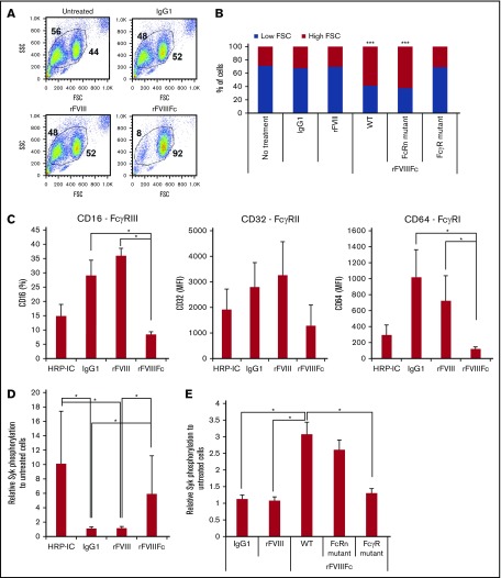
rFVIIIFc induces Fcγ receptor internalization and signaling in an Fc-dependent fashion. (A) Forward scatter (FSC)/side scatter (SSC) pseudocolor plots of macrophages treated with IgG1, rFVIII, or rFVIIIFc (each at 200 nM) for 24 hours, illustrating the size change of the cells upon rFVIIIFc treatment. Numbers in FSC/SSC pseudocolor plots indicate the percentage of smaller and larger cell populations. One representative experiment is shown. (B) Graph shows cumulative data on macrophage size based on the percentages of low-FSC and high-FSC cell populations, observed by flow cytometry (n = 12). Significance indicated compared with untreated cells. (C) Macrophages (n = 4) were treated with HRP-ICs as positive control, IgG1 as negative control, rFVIII, or rFVIIIFc for 24 hours, and the cell surface expression of the FcγRs CD16, CD32, and CD64 was measured by flow cytometry. Downregulation of the FcγRs from the cell surface indicates internalization. (D) Macrophages (n = 10) were treated with HRP-IC, IgG1, rFVIII, or rFVIIIFc for 15 minutes, and the phosphorylation of Syk was measured using the Meso Scale Diagnostics (MSD) platform. Upregulation of pSyk levels shows direct FcγR engagement by HRP-IC and rFVIIIFc. (E) Mutant rFVIIIFc molecules unable to bind the FcRn (FcRn mutant) or the FcγRs (FcγR mutant) were used to treat macrophages (n = 4) along with the wild-type rFVIIIFc (WT), IgG1, and rFVIII for 30 minutes, and Syk phosphorylation was measured using the MSD platform. Downregulation of pSyk after FcRn and FcγR mutant treatment compared with WT rFVIIIFc shows the role of these receptors in signal transduction. Mean ± SE; *P ≤ .05, ***P ≤ .005.

