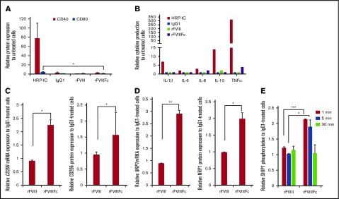
Figure 2.
rFVIIIFc treatment does not activate macrophages. (A) Macrophages (n = 3) were treated with IgG1, rFVIII, or rFVIIIFc for 24 hours, and the cell surface expression of the costimulatory/activation molecules CD40 and CD80 were measured by flow cytometry. The absence of significant molecule upregulation shows no classical activation of macrophages upon rFVIIIFc treatment. (B) Macrophages (n = 8) were treated with HRP-IC, IgG1, rFVIII, or rFVIIIFc for 24 hours, and the production of interleukin-1β (IL-1β), IL-6, IL-8, IL-10, and tumor necrosis factor α (TNFα) cytokines were measured from cell supernatants using multiplex enzyme-linked immunosorbent assay. Compared with the proinflammatory activation signal from HRP-IC, rFVIIIFc treatment of macrophages did not significantly upregulate proinflammatory cytokine production (error bars and significance not shown for graph clarity). RNA and protein levels of the alternatively activated macrophage markers mannose receptor CD206 (n = 7) (C) and NRP1 (n = 3) (D) were measured by quantitative PCR (qPCR) and flow cytometry, respectively. (E) Macrophages (n = 3) were treated with IgG1, rFVIII, or rFVIIIFc for 1, 5, or 30 minutes, and phosphorylated SHIP1 was measured using the MSD platform. Significant elevation of pSHIP1 levels after rFVIIIFc treatment compared with IgG1 or rFVIII treatments shows triggering of inhibitory signaling events. Mean ± SE; *P ≤ .05, **P ≤ .01, ***P ≤ .005.

