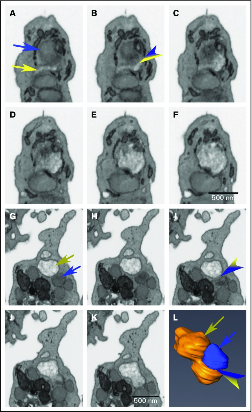
Figure 5.
α-Granule matrix decondensation propagates from open canalicular system–granule fusion or decondensed-condensed granule fusion sites. Images are individual slices from FIB-SEM image stacks, and the frames shown are spaced 15 nm apart. (A-F) The blue arrow indicates a condensed α-granule, and the yellow indicates open canalicular system–elements (A). The variegated blue/yellow arrowhead indicates a CS-granule fusion zone (B). The dark tubular structures adhering to the decondensing α-granule are elements of dense tubular network. (G-L) The tan arrow indicates a decondensed α-granule (G), and the blue arrow indicates a condensed α-granule (G). The variegated tan/blue arrowhead indicates the decondensed-condensed granule fusion zone in the FIB-SEM slice (I) and similarly marks the fusion zone in a rendering (L) that is slightly tilted from the perpendicular relative to the FIB-SEM image planes. Bars represent 0.5 μm.

