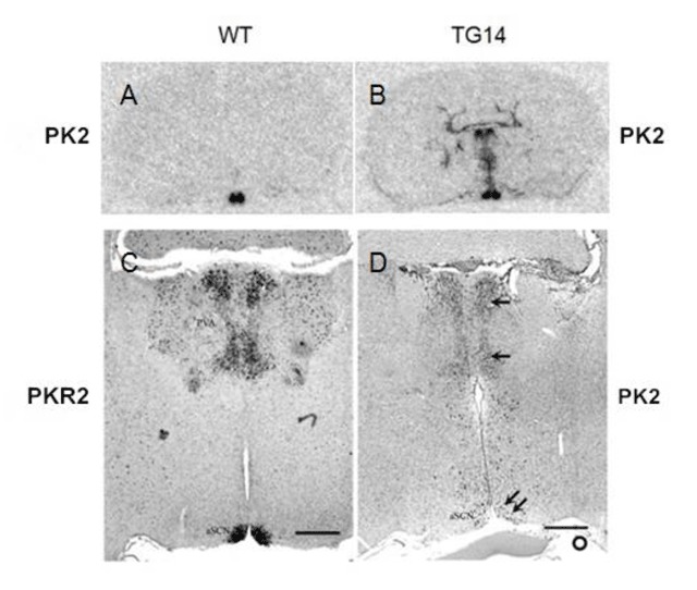
Figure 1.

Expression of PK2 in the brain sections of the PKR2-PK2 transgenic mice. PK2 or PKR2 that are labeled at the left and right of the images are the probes used in the in situ hybridization. A and B show the expression of PK2 by in situ hybridization with 35S probe on brain sections that were sampled at ZT4. Note the selective expression of PK2 in the wild type mice (A) and the ectopic expression of PK2 in transgenic mice, in addition to its expression in the SCN (B). C and D show the in situ hybridization with digoxigenin-labelled probes. The ectopic expression of PK2 in the transgenic mice (single arrow, D) was apparent in the midline thalamus. Such ectopic expression of PK2 correlates with the expression of PKR2 (C). PK2 mRNA was detected in the SCN of the transgenic mouse sampled at ZT18 (double arrow, panel D). At ZT18, only very low level of PK2 expression was found in the SCN of wild type mice. Scale bar, 500 μm.
