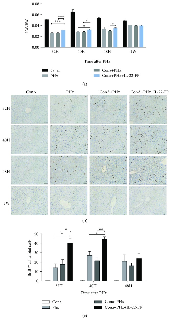
Figure 2.
IL-22-FP enhances liver regeneration in a ConA model after PHx. C57BL/6 mice (8 to 10 wk old) were randomly divided into 4 groups: the ConA group, PHx group, ConA + PHx group, and ConA + PHx + IL-22-FP group. Each group of mice was treated as described in Materials and Methods. (a) The liver weight/body weight ratio (LW/BW) of each group at 32 h, 40 h, 48 h, and 1 wk post-PHx; n = 4 for each group. (b) Bromodeoxyuridine (BrdU) staining of mouse liver at 32 h, 40 h, 48 h, and 1 wk post-PHx (400x magnification). (c) BrdU+ hepatocyte/total hepatocyte ratios of 4 groups of mice at 32, 40, and 48 h post-PHx. Pictures were taken at 400x magnification; n = 4 for each group. ∗p < 0.05, ∗∗p < 0.01, and ∗∗∗p < 0.001.

