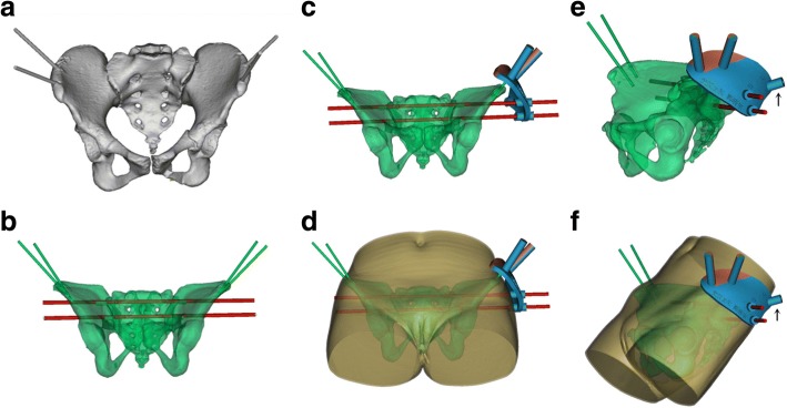
Fig. 3.
External template designed using Mimics software. a A 3D model of the pelvis was reconstructed including the marker pins. b S1 and S2 virtual screws were placed into the sacrum and adjusted to the midway of the osseous corridor without any penetration. c-f The template was designed to connect the marker pins and virtual screws, providing sleeves to attach the template on the marker pins and guide the K-wire to the target corridor. Black arrow indicates the guide sleeve for the anterior column screw. (d/f) The plate is low-profile, minimizing the distance between both template and skin, and marker pins and virtual screws

