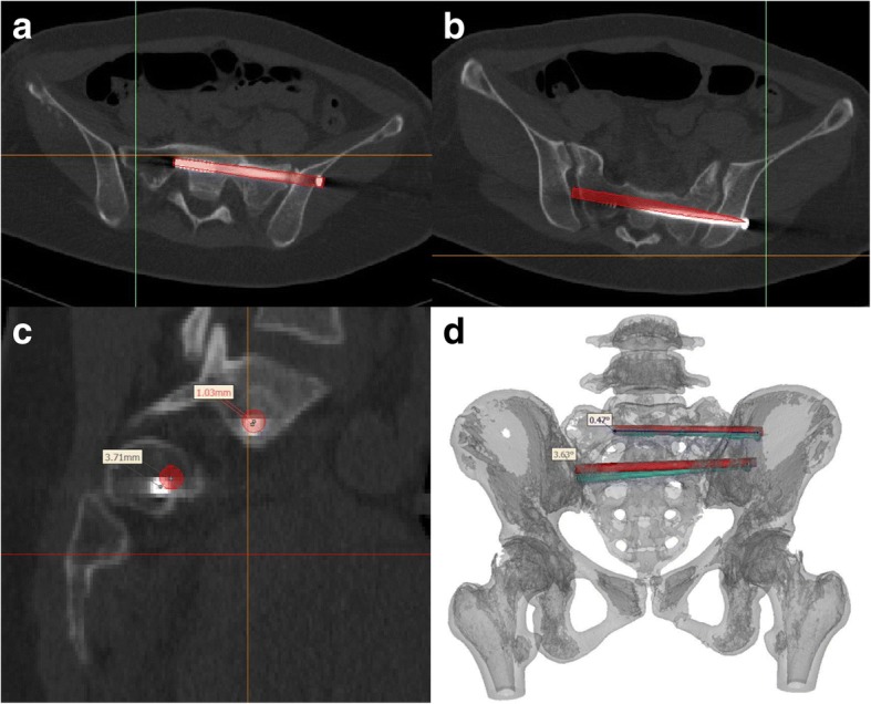
Fig. 5.

The procedure for postoperative measurements. a-b The S1 and S2 axial views obtained after insertion of the partially threaded screws were merged with the pre-operative images used for planning (red bar). c The deviation distance between the inserted and planned virtual screw was measured on the sagittal plane at the nerve root tunnel zone. d The deviation angle was measured on the superimposed image of the pre- and postoperative 3D reconstructions
