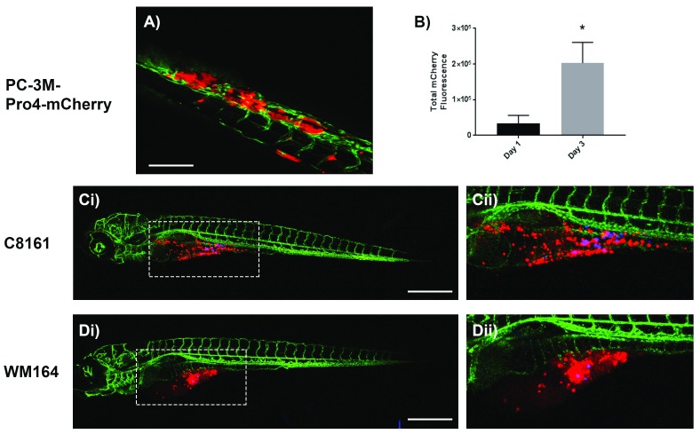
Figure 3. Representative confocal z-stack images of kdrl-GFP casper zebrafish embryos 72 hours after injection with human cancer cells.
A) PC-3M-Pro4-mCherry prostate cancer cells injected into the duct of Cuvier form tumours in the caudal hematopoietic tissue of the zebrafish tail; Scale bar = 150 μm. B) Quantification of total mCherry fluorescence by prostate cancer cells after 1 and 3 days post injection; n=4, *p<0.01, 0.05 CI, paired t-test. Ci– ii) C8161 and Di– ii) WM164 melanoma cells (stained with Red DiI dye) injected alongside FluoSpheres (Blue) into the yolk sac survive and invade throughout the yolk sac; Scale bar = 500 μm.

