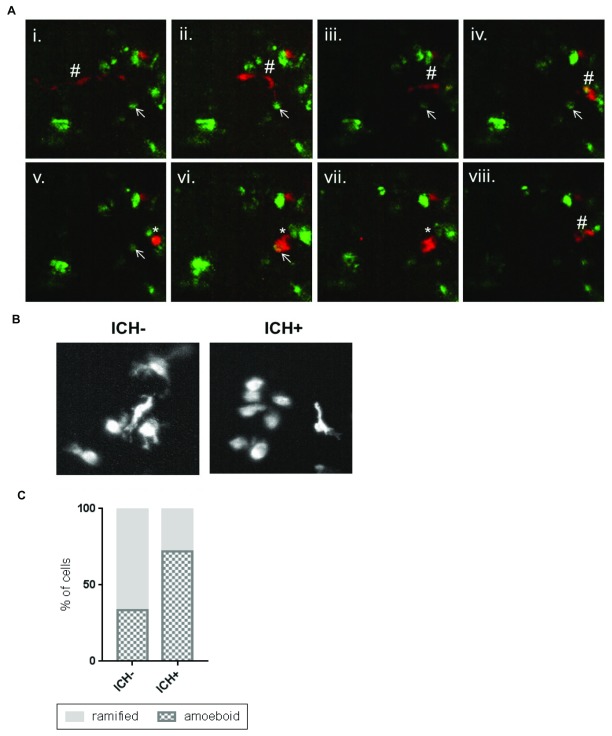
Figure 5. Activated macrophage cells show a phagocytic response to the brain lesion.
( A) Representative time-lapse stills (from Supplementary Video 1) showing a ramified patrolling macrophage migrating towards an annexinV positive cell (i - vi). The macrophage acquired an amoeboid morphology (v) before phagocytosing the annexinV-positive cell (vi, vii). After phagocytosis the macrophage resumes a ramified morphology and migrates away and the annexinV-positive cell can no longer be seen (viii). Ramified macrophage (#), annexinV positive cell (arrow), amoeboid macrophage (*). ( B) Representative images of mpeg1-positive cells in the intracerebral haemorrhage (ICH)- and ICH+ larval brain exhibiting amoeboid and ramified morphologies. ( C) An increased proportion of amoeboid (phagocytic) and decreased proportion of ramified (inactive) macrophages was observed in ICH+ brains in comparison to ICH- siblings.

