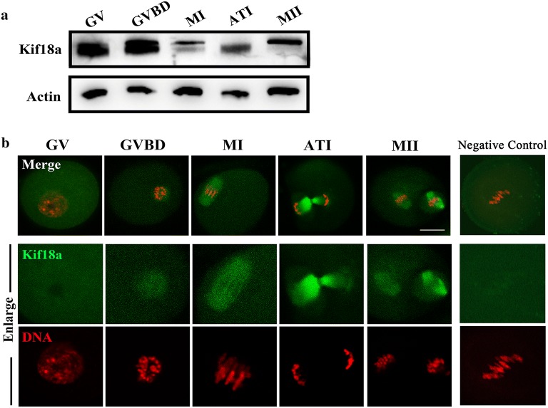
Fig. 1.
Expression and localization of Kif18a in mouse oocytes. a Oocytes at different maturation stages were examined by Western blotting. b Oocytes from GV to MII stages were stained with anti-Kif18a antibody (green) and counterstained with DAPI for DNA visualization (red). After GVBD, Kif18a accumulated around chromosomes. Also, Kif18a localized in the meiotic spindle region at both the MI and MII stages, while it localized in the midbody at the ATI stage. Negative control was stained with secondary antibody without Kif18a. Scale bar: 20 μm

