Fig. 2.

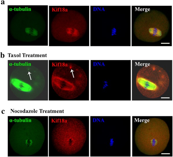
Kif18 localization after taxol or nocodazole treatment. a Double staining of MI oocytes with an anti-Kif18a antibody (red) and an anti-α-tubulin antibody (green). Oocytes were counterstained with DAPI to visualize DNA (blue). Kif18a mainly localized on the meiotic spindle. b Subcellular localization of Kif18a after taxol treatment during mouse oocyte meiotic maturation. Arrows indicated asters. Green, α-tubulin; red, Kif18a. c Subcellular localization of Kif18a after nocodazole treatment during mouse oocyte meiotic maturation. Green, α-tubulin; red, Kif18a. Scale bar: 20 μm
