
Fig. 1.
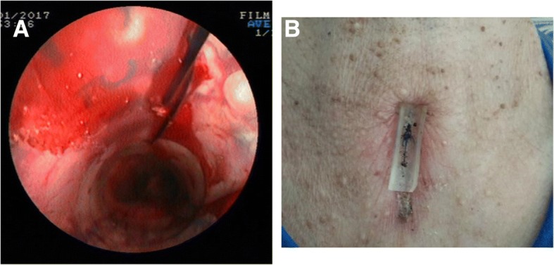
Images of the operation procedures. a: Suture fixation method for the tracheal stent. b: Postoperative CT showing the silicon pad on the skin at the puncture site (as the arrow indicates)
Official websites use .gov
A
.gov website belongs to an official
government organization in the United States.
Secure .gov websites use HTTPS
A lock (
) or https:// means you've safely
connected to the .gov website. Share sensitive
information only on official, secure websites.
Images of the operation procedures. a: Suture fixation method for the tracheal stent. b: Postoperative CT showing the silicon pad on the skin at the puncture site (as the arrow indicates)