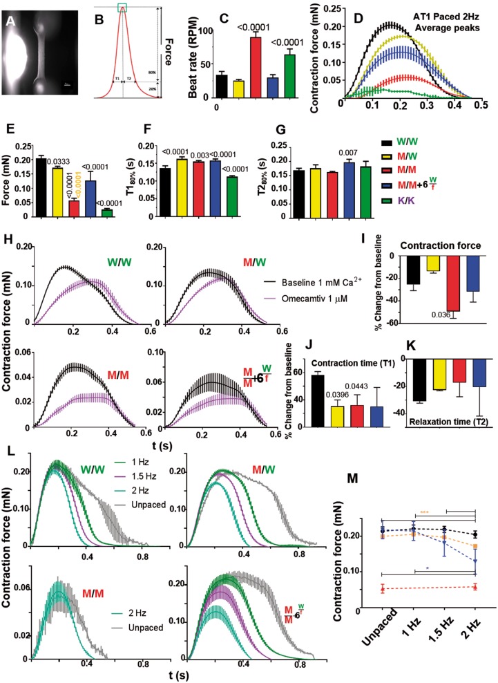
Figure 6.
Contractile force analysis in AT1-human engineered heart tissues. (A) Fibrin-based AT1-human engineered heart tissue attached to silicone posts (Bar = 1 mm). (B) Schematic contraction peak showing parameters analysed, providing data on (C) spontaneous beat rate (n = 8). Electrically paced engineered heart tissues produced average contraction peaks (D), quantified for (E) contraction force, (F) contraction time, and (G) relaxation time (n = 4). (H) 2 Hz electrically paced AT1-engineered heart tissues with or without omecamtiv mecarbil treatment produced average contraction peaks, quantified for (I) contraction force, (J) contraction time, and (K) relaxation time (n = 3). (L) Force-frequency relationship in MYH7-mutant AT1-engineered heart tissues was assessed and quantified in (M). Fast spontaneous beat rate of homozygous R453C-β-myosin heavy chain mutant meant only 2 Hz pacing was possible. (n = 4). Data, mean ± SD. P-values, one-way ANOVA test + Dunnett’s correction.

